For decades, a Parkinson’s diagnosis came with a relatively clear biological story. That story is getting a lot more complicated, and for patients, the implications could be significant. A growing body of research suggests that for many Parkinson’s patients, the disease they were diagnosed with may only be part of the story; it’s not a one-stop shop anymore.
In many Parkinson’s patients, the brain isn’t just dealing with Parkinson’s. It’s also accumulating the same toxic proteins you’d find in people with Alzheimer’s or even ALS (Lou Gehrig’s disease).
Your body makes certain proteins that are supposed to help your cells function. But sometimes those proteins get misshapen. A protein has to fold into a very specific shape to do its job. When it folds wrong, it’s useless at best, and at worst, it starts sticking to other misfolded proteins, building up into clumps your brain can’t easily break down. When that happens, other systems in your body are impacted, and symptoms start to appear.
There is a growing consensus among researchers and neurologists that Parkinson’s is not a straightforward condition, but a combination of several overlapping disease processes taking place at the same time, some that are associated with other neurological diseases. These include Alpha-synuclein, the main protein associated with Parkinson’s disease; Amyloid-beta plaques and Tau tangles, both hallmarks of Alzheimer’s disease; and TDP-43, the protein considered the biological hallmark of ALS (Amyotrophic lateral sclerosis).
What these pathologies all have in common is that the proteins involved become misfolded and clump together. In the case of Alpha-synuclein, those clumps form what are known as Lewy bodies, which are toxic to surrounding tissue. Once these clumps develop, neurons struggle to send and receive the electrical signals they rely on to communicate. The brain’s own waste-removal system, known as the Glymphatic System, also becomes impaired — overwhelmed by the volume of toxic proteins and unable to efficiently clear harmful buildup from the brain.
To understand the significance of TDP-43 in the context of Parkinson’s, it’s important to understand how TDP-43 relates to ALS. In ALS patients, misfolded TDP-43 is so consistently present that researchers consider it the hallmark of the disease. The protein attacks the nerve cells responsible for controlling voluntary movement, killing off the motor neurons responsible for communication between the brain and the muscles. People with the disease progressively lose their mobility, and the disease is fatal — the average lifespan after diagnosis is around three years, though others have lived longer. Researchers continue to work on an explanation for the cause, but aging, cellular stress, and genetic mutations are all considered contributing factors.
Several papers have been published on the presence of TDP-43 in Parkinson’s patients, with studies showing that TDP-43-dependent missplicing is widespread in the disease. One 2009 study found TDP-43 pathology in roughly 7% of Parkinson’s cases, while studies focusing on Parkinson’s disease dementia (PDD) report the figure climbing to around 19%. In a review titled Co- and Multi-Pathologies in Parkinson’s Disease: An International Parkinson and Movement Disorder Society Scientific Issues Committee Review, the researchers have found aggregates of TDP-43 in areas of the brain that are central to memory, learning, emotional processing, and cognition. The proteins are believed to disrupt communication between cells, disrupt neuron function, and contribute to dementia symptoms. The effects of the TDP-43 pathology appear more frequently in Parkinson’s patients who develop dementia than in those who don’t, suggesting TDP-43 can impact as disease progression continues, spreading into the cortical regions, memory systems, and limbic structures of the brain, the areas that govern thought, memory, and emotion.
The job of TDP-43 is to regulate RNA, oversee protein production, and help with the maintenance of the cell, but when the protein shape becomes abnormal, it abandons the cell’s nucleus, misfolds, and aggregates, impairing protein production, disrupting neuron communication, and ultimately contributing to cell death. The brain, in effect, loses part of its internal management system. So, for Parkinson’s patients, finding TDP-43 in the substantia nigra, the dopamine-producing region most heavily damaged in Parkinson’s, contributes to the loss of motor function even in the absence of Lewy bodies.
The review makes clear that Parkinson’s is far more complex than a single biological explanation can account for, not purely α-synuclein or a single disease entity. When it comes to TDP-43: Rather than acting as a single toxic protein, researchers increasingly suspect TDP-43 may contribute to a broader breakdown of essential cellular maintenance systems inside neurons, with multiple overlapping biological systems contributing to Parkinson’s disease.
Researchers are also trying to understand why some areas of the brain are hit harder than others. Regions like the substantia nigra, hippocampus, and motor neurons show some of the earliest damage, with researchers believing they carry heavy energy loads, maintain long communication networks throughout the body, and are particularly vulnerable to oxidative stress, inflammation, and mitochondrial strain. The combination of that much demand and vulnerability may make them the first to break down when something goes wrong, much like parts on a car engine. What is clear is that our understanding of Parkinson’s is evolving — it’s a disease that can’t be understood under a single scope. The presence of TDP-43, alongside the pathologies of Alzheimer’s and ALS, suggests that the boundaries between these diseases may be far less defined than once believed. For researchers, that opportunity is enormous, one that may lead not only to better treatments for Parkinson’s, but for other neurodegenerative diseases as well.

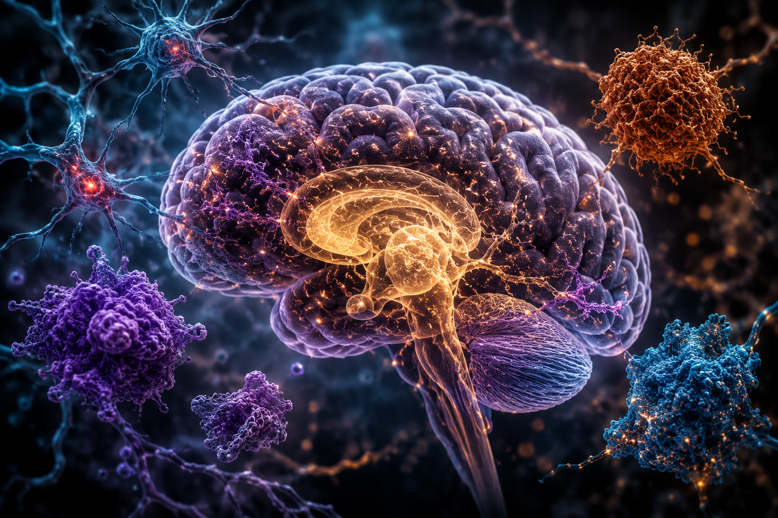
Image by Chris Denny/ChatGPT
Discover more from
Subscribe to get the latest posts sent to your email.